Ultrasonography (also called ultrasound or sonography) is a noninvasive, pain-free procedure that uses sound waves to examine a pet’s internal organs and other structures inside the body. It can be used to evaluate the animal’s heart, kidneys, liver, gallbladder, and bladder; to detect fluid, cysts, tumors, or abscesses; and to confirm pregnancy or monitor an ongoing pregnancy.
We may use this imaging technique in conjunction with radiography (x-rays) and other diagnostic methods to ensure a proper diagnosis. Interpretation of ultrasound images requires great skill on the part of the clinician.
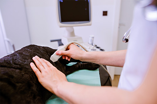
The ultrasonographer applies gel to the surface of the body and then methodically moves a transducer (a small handheld tool) across the skin to record images of the area of interest. The gel helps the transducer slide more easily and creates a more accurate visual image.
The transducer emits ultrasonic sound waves, which are directed into the body toward the structures to be examined. The waves create echoes of varying degrees depending on the density of the tissue and the amount of fluid present. Those waves create detailed images of the structures, which are shown on a monitor and recorded for evaluation.
Ultrasound does not involve radiation, has no known side effects, and doesn’t typically require pets to be sedated or anesthetized. The hair in the area to be examined usually needs to be shaved so the ultrasonographer can obtain a good result.
If you have any questions about our ultrasonography service or what to expect during your pet’s procedure, please don’t hesitate to ask.
Veterinary ultrasound was originally used to diagnose a pregnancy. Today it is used for a much wider range of information. An ultrasound can give a visualization of the inside of your pet’s abdomen. It allows the trained technician to view internal organs and structures such as the liver, spleen, kidneys, bladder and intestinal tract. We have Veterinary Technicians who have completed training by Oncura Partners to perform abdominal ultrasounds. During the ultrasound, they can request that an expert sonographer gain remote access to further direct them during the ultrasound. This will allow the best possible pictures for evaluation. The pictures are then read and interpreted by a board-certified specialist. This information, together with laboratory results, can help the doctor to prescribe the best treatment for your pet possible.